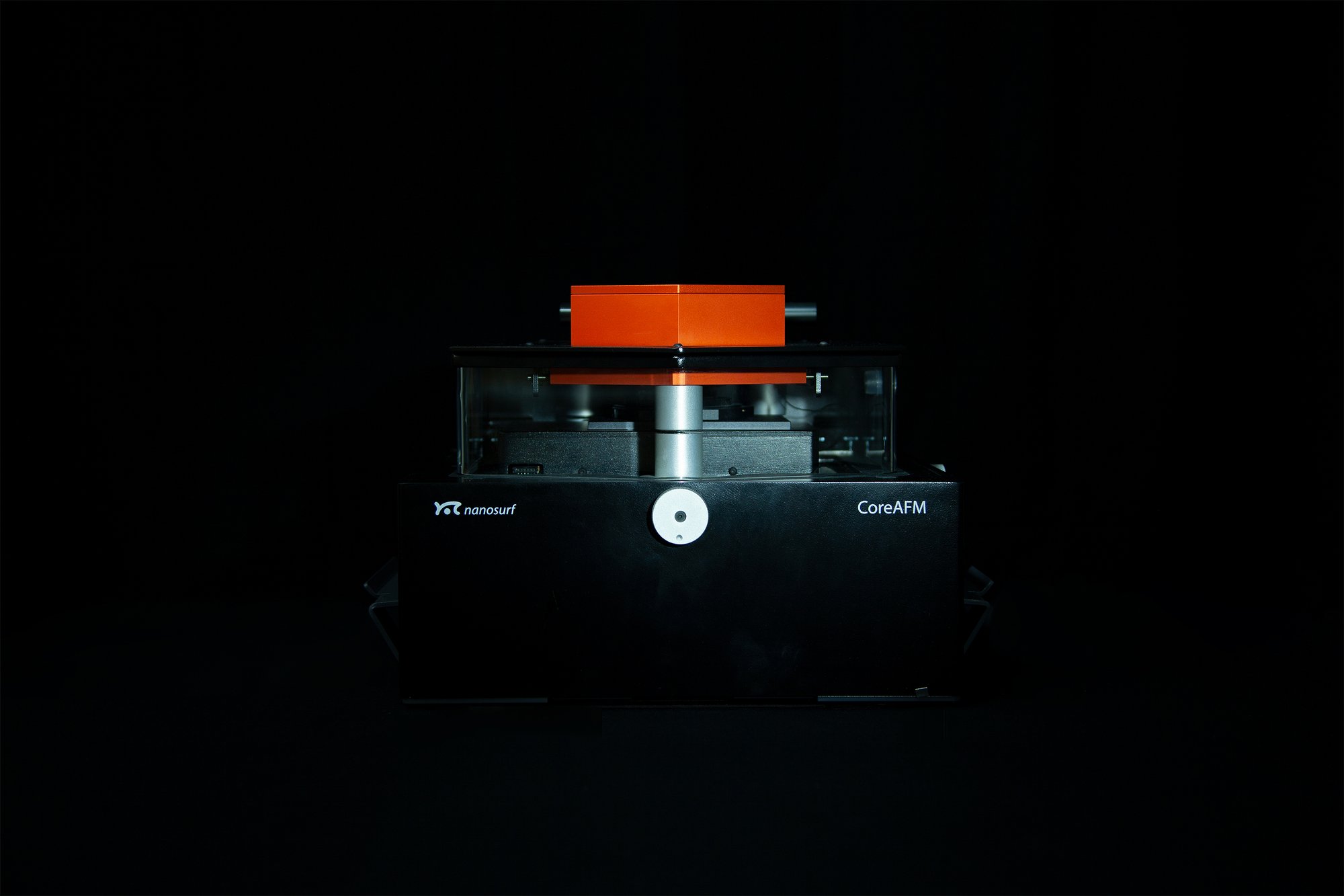
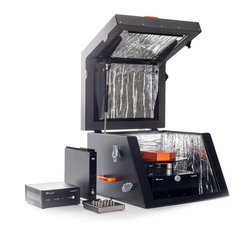
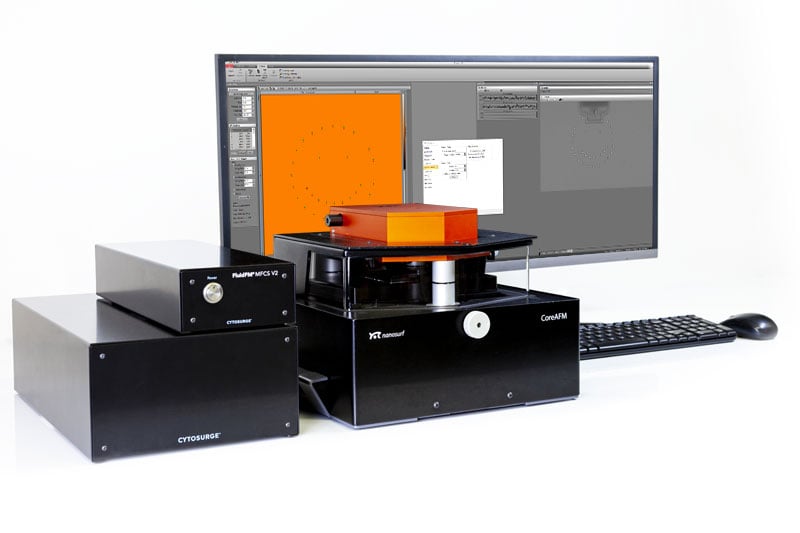
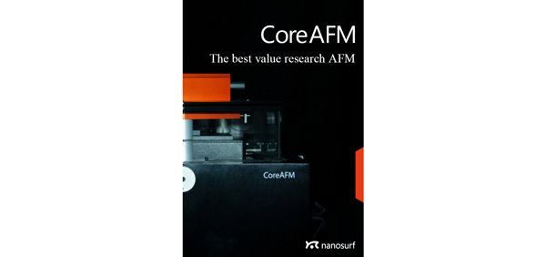
CoreAFM
The Best Value Research AFM
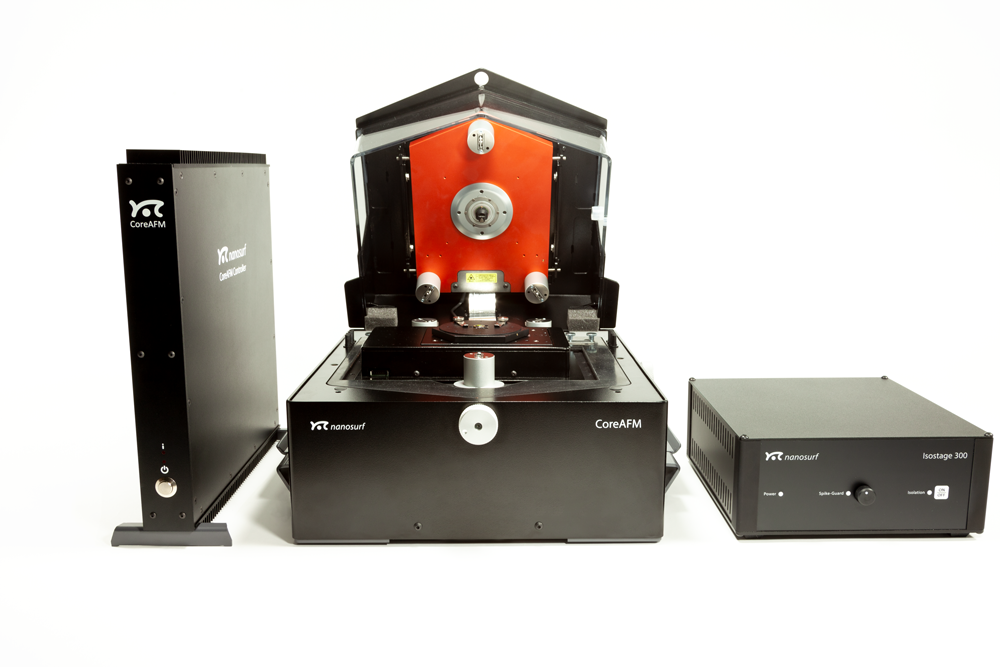
The CoreAFM was designed to incorporate all the essential elements of an AFM in an easy to use, integrated single unit: a modern flexure-guided scanner, an XYZ sample stage, a camera for sample observation, an active vibration table, and an airflow shielding. The specially designed 24-bit controller ensures precise and reliable control over the scanning process, as well as easy data acquisition and analysis. The CoreAFM system has everything you need to start exploring the nanoworld with AFM; just connect the controller, plug in power and USB, and you are ready to go.
Speak with an expert
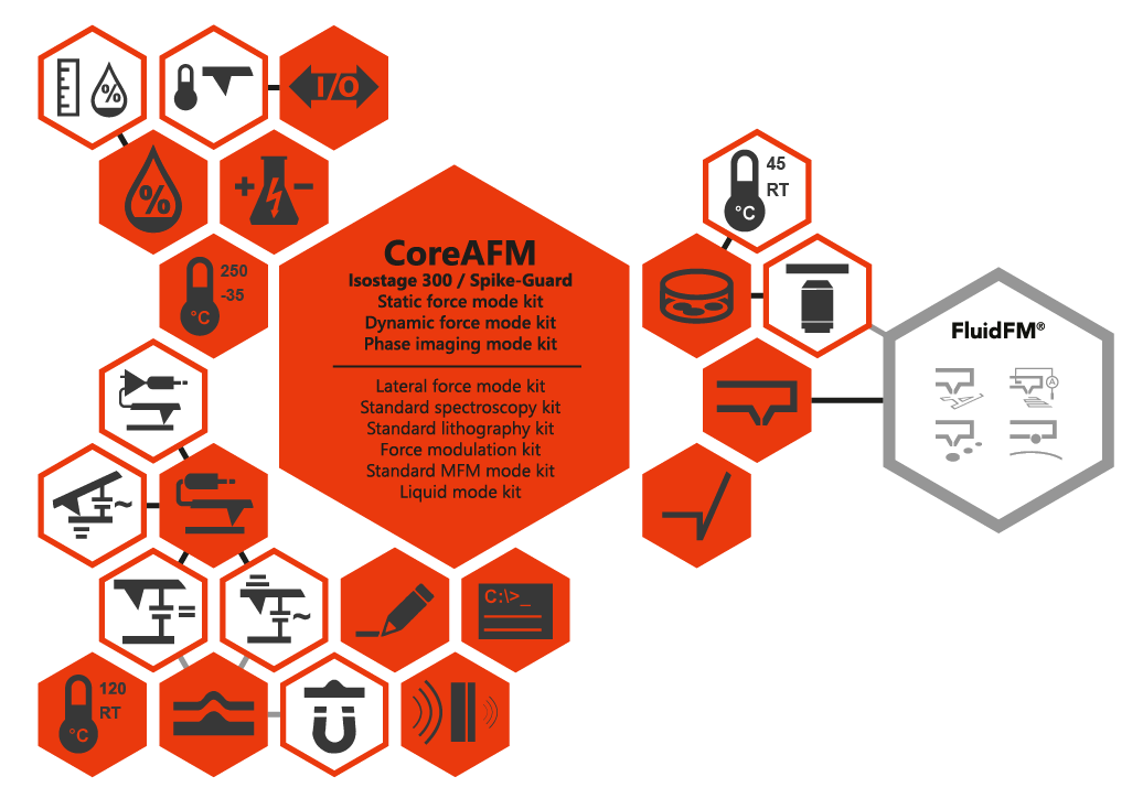
Seamlessly extendable functionality
The CoreAFM is a versatile and powerful instrument that can handle various applications in both air and liquid environments. Whether you need to perform basic or advanced measurements, the CoreAFM adapts to your needs with its modular design and more than 30 optional modes and functions. Advanced options include sample heating, environmental control, in-plane magnetic field control, and scripting. The CoreAFM is more than just an all-in-one atomic force microscope, it is a complete solution for your nanoscale research and analysis.
Basic research AFM with a competitive price tag
If you are looking for a reliable and versatile instrument for basic research, the CoreAFM is the right choice for you. This system offers a solid foundation for a multitude of applications in nanoscience and nanotechnology, with the option to expand the feature set as your research evolves. The CoreAFM is a cost-effective solution that delivers high quality without compromising your budget. Whether it is to explore surface morphology or study electrical phenomena, the CoreAFM will help you achieve your objectives with ease and accuracy.
CoreAFM for Materials Research
CoreAFM incorporates all the essential elements of a research AFM in an easy to use adaptable platform. The precision and performance are well-suited for a wide variety of applications in materials science and related fields. Powerful imaging and precise electrical characterization modes provide you with a robust and versatile research instrument that shines under the pressure of a tight budget.
Precision and Versatility
Precision and Versatility
The CoreAFM is a powerful and versatile instrument that can handle a wide range of applications in material science and beyond. Whether you are working with polymers, ceramics, metals or 2D materials, the CoreAFM provides you with high-quality data and insights. With more than 30 modes and functionalities to choose from, the CoreAFM allows you to explore different aspects of your samples, such as morphology, mechanical properties, electrical properties and more.
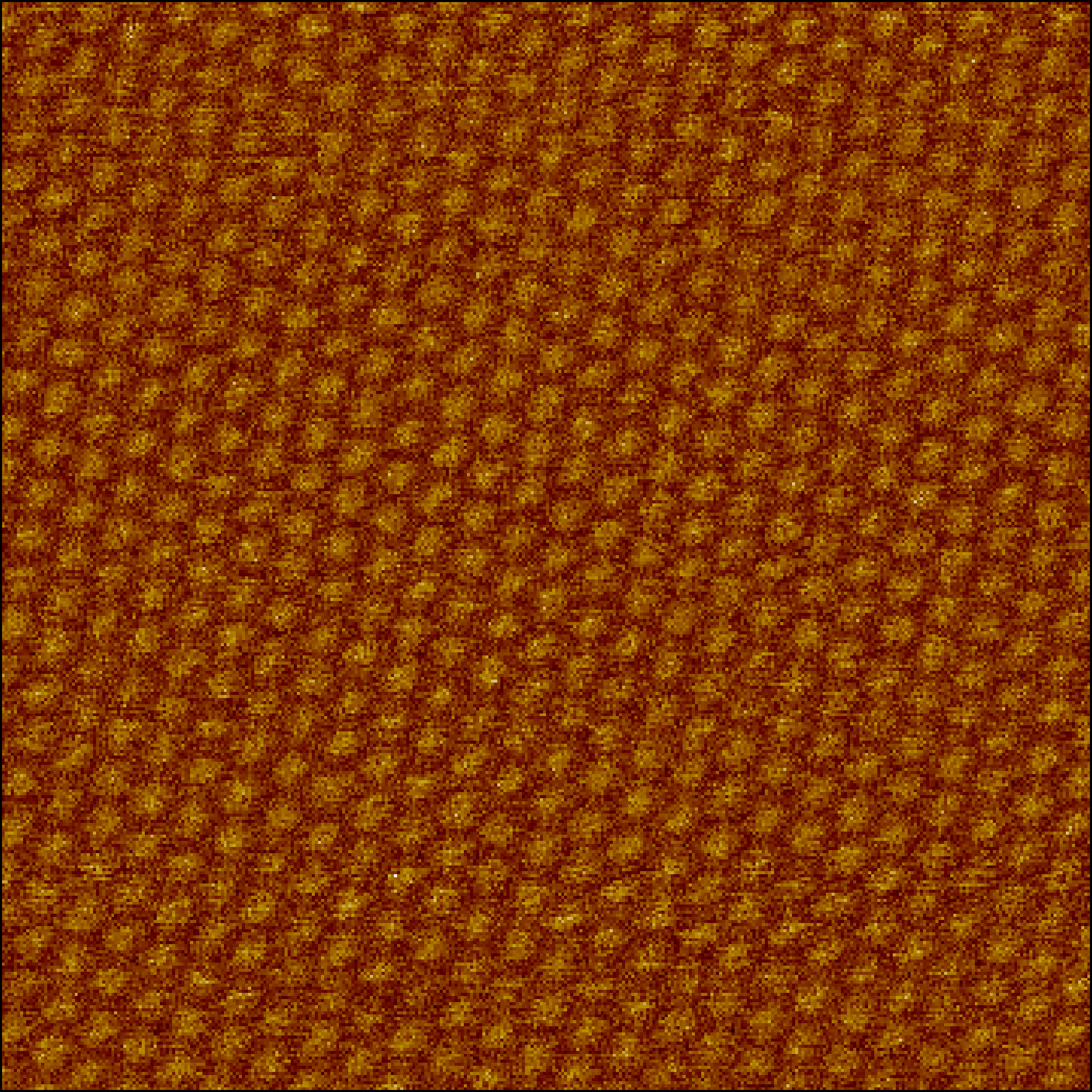
Image: Moiré superlattice of twisted graphene on hBN; image courtesy Nanoelectronics group TIFR, India.
Nano-Electrical Characterization
Nano-Electrical Characterization
With the CoreAFM, you can explore the electrical properties of various materials with unprecedented precision and versatility. Whether you are interested in the conductivity, capacitance, magnetism, or piezoelectricity of your samples, the CoreAFM has a mode for you. From C-AFM to SMM, the CoreAFM covers a wide range of electrical AFM techniques that can reveal the nanoscale secrets of 2D materials, metals, piezo-active materials and semiconductors. The CoreAFM is not just another AFM instrument, it is a powerful tool for electrical nanoscience.
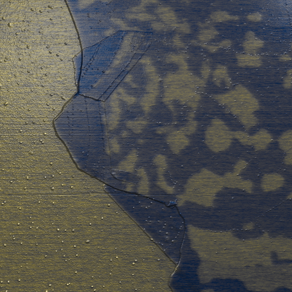
Image: Contact potential difference on multilayer graphene flake
Effective and Affordable
Effective and Affordable
The CoreAFM combines precision and affordability, making it a smart choice for your research needs. Configure the functionality of your system to perfectly meet your needs, and purchase only what you need. The base configuration includes everything vital for an AFM to perform effectively, and the universe of functionality options offers everything you need to tailor a lean system that supports your research perfectly.
CoreAFM for Life Science
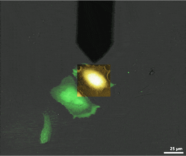
CoreAFM does not look like the typical bio-AFM, but thanks to its smart engineering can perform very well in certain areas of life-science research. The functionality set includes all features commonly required in life science focused AFM, and the vital aspect of microscope integration is available through the DIMO. CoreAFM is the only AFM in its class with versatility covering even biological applications. It’s also the most affordable path to FluidFM functionality.
FluidFM
FluidFM
FluidFM is a revolutionary technology that combines microfluidics and atomic force microscopy to enable precise manipulation, deposition and analysis of nanoscale objects and single cells. The CoreAFM is a versatile and user-friendly AFM system that supports FluidFM integration and software control. With FluidFM, you can perform a wide range of applications such as nanolithography and nanoinjection as well as single-cell extraction and force spectroscopy.

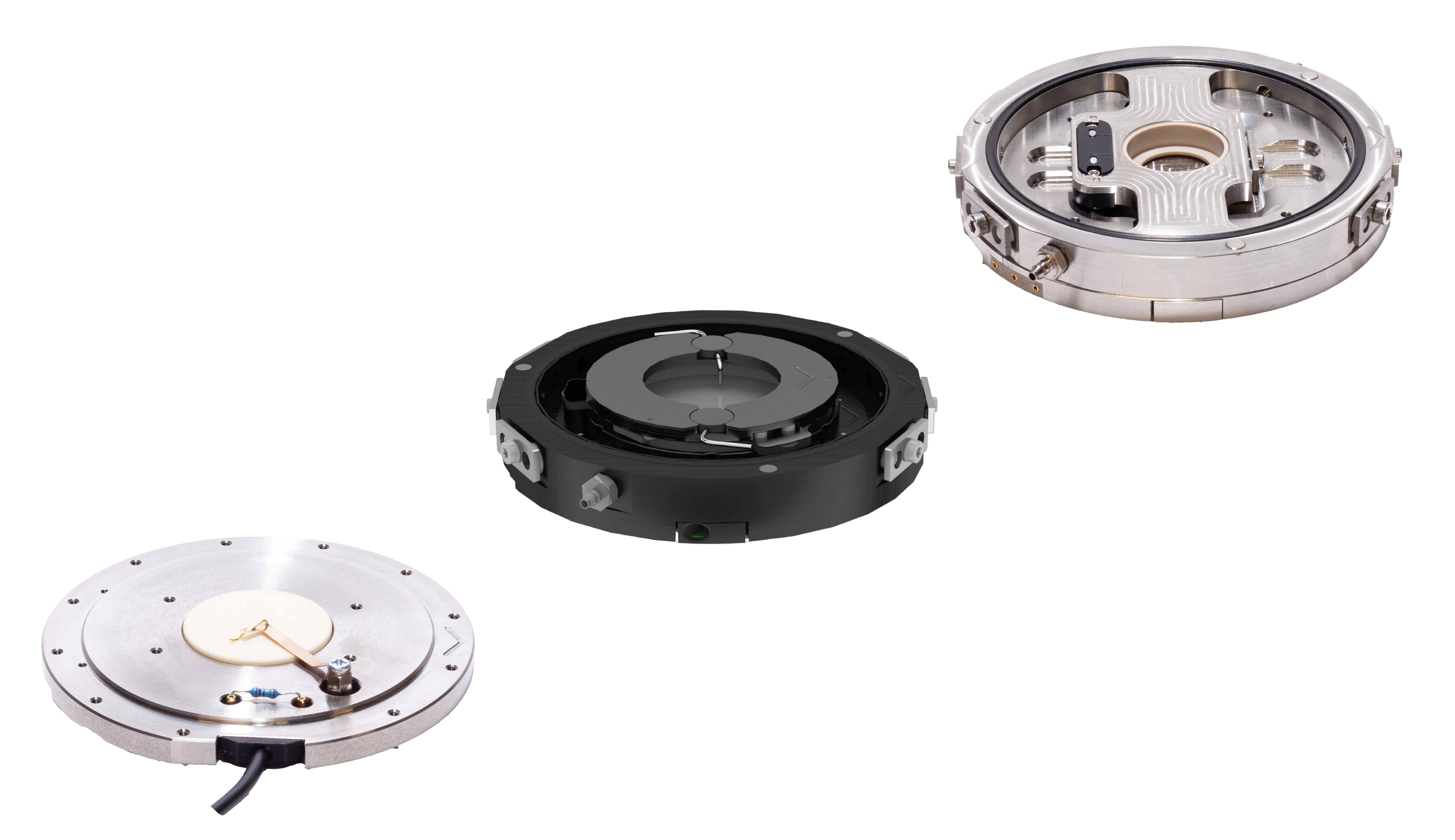
DIMO
DIMO
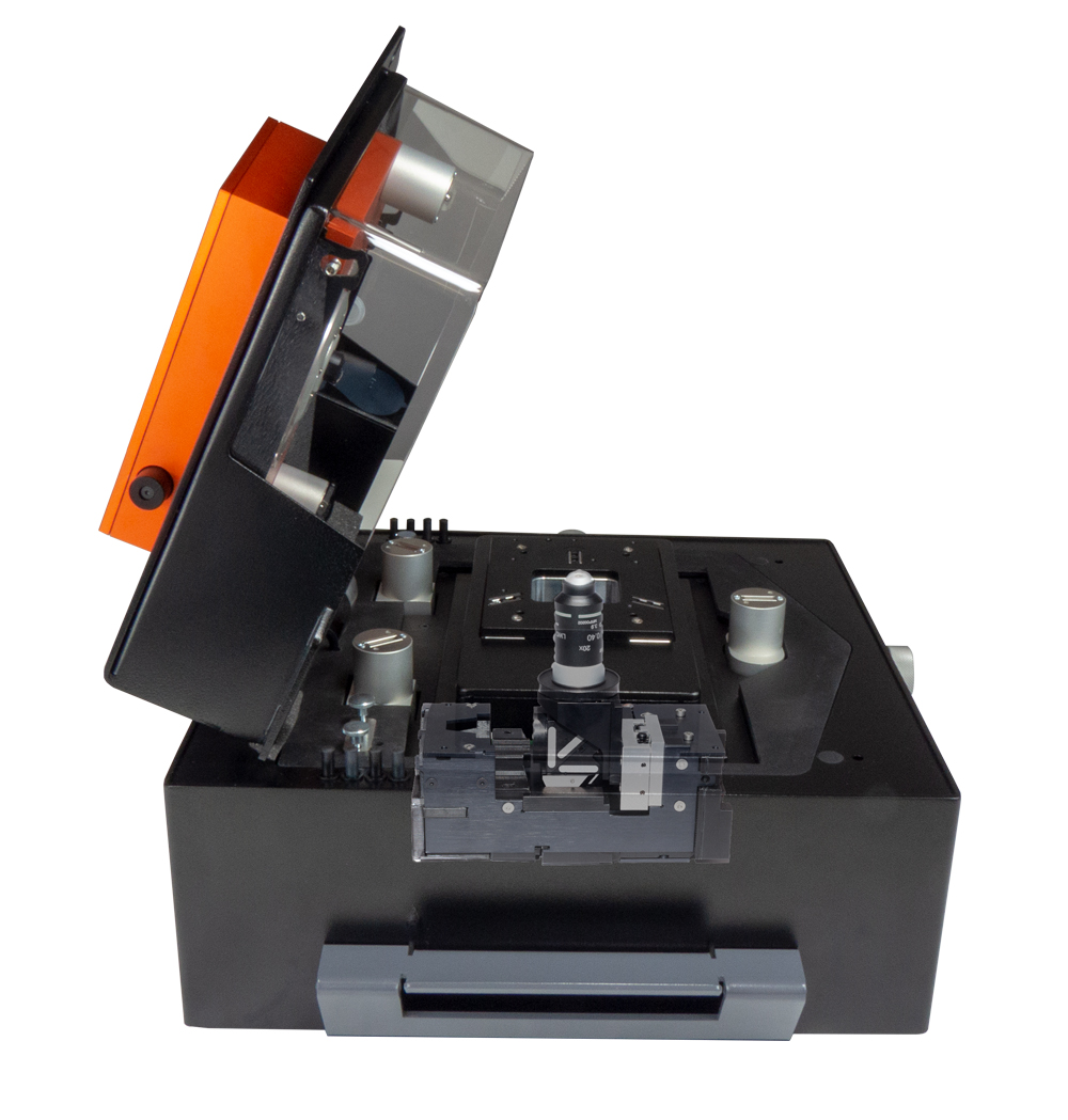
The Digital Inverted Microscope Option (DIMO) combines the best of both worlds: the compact footprint of a stand-alone AFM system and the capability to observe samples from below. It allows you to view your samples with transmitted light or by fluorescence imaging. The DIMO also enables FluidFM applications on small systems, giving you more flexibility, control and versatility in your research. The DIMO features high-resolution optics with interchangeable objectives, so you can adjust the magnification and resolution to suit your needs.
Bio Functionality
Bio Functionality
From temperature control to Petri-dish sample holder and 150 µm z actuator, the CoreAFM has all the functionality a well-rounded AFM needs to deliver results in biological research. If you value versatility and accessibility over focused specialization, the CoreAFM is what you’ve been searching for, especially when under budget constraints.
Dr. Joshua D. McGraw,
CNRS ESPCI-PSL / IPGG
In my lab at ESPCI, we study the interfacial dynamics of soft materials. Usually, a polymeric material makes up either the surrounding fluid or a surface layer and a colloid can be used as a tracer or model object interacting with the surface. As such, AFM in general, and colloidal probe AFM in diverse environments more specifically, is a necessity for my lab. With such applications in mind, and with a young researcher’s budget, Marco from Nanosurf’s technical sales team suggested the CoreAFM.
In addition to performing very well in air for surface topographic characterization, the instrument also performs under liquid environments and at elevated temperatures using a colloidal probe. This latter functionality is possible thanks to theCoreAFM’s impressive modularity. An extensive library of accessories and functional add-ons, all with a good value for money, give me the flexibility to upgrade the instrument to suit my research needs as they arise. Lastly, the technical team at Nanosurf is very available for troubleshooting support when the need arises, and have always helped to solve problems efficiently. Given the instrument’s value-for-money, the personal and efficient technical support, the instrument’s performance and modularity, I would not hesitate to recommend the CoreAFM.
Download the CoreAFM Brochure
The PDF brochure includes details on the CoreAFM's technology, application examples for different areas of interest and system specifications.
#{ item.resourceType }
#{ item.date_text_field }
#{ item.name }
#{ truncateText(item.metadescription) }
#{ item.readmoretext }Example Measurements
#{ row.name }
There are no items to display.
#{ item.resourceType }
#{ item.date_text_field }
#{ item.name }
#{ truncateText(item.metadescription) }
#{ item.readmoretext }Interested in CoreAFM?
Get to know the CoreAFM! Reach out to us to discuss your application with one of our AFM experts, discuss your needs for a budgetary quote or schedule a product demonstration or exploratory meeting.