Discover the capabilities of WaveMode AFM in characterizing bottlebrush polymers with unprecedented detail and speed, ...

05.04.2024

Héctor here, your AFM expert at Nanosurf calling out for people to share their Friday afternoon experiments. Today I expand my knowledge on the structure of shells.
Why shells?
Why shells are a recurring theme on fridayAFM?
Because shells are light, easy to tool, long lasting biological structures with interesting micrometer-scale substructures. So if we can learn something about the microstructure, maybe we can figure out how to make materials with similar properties.
What have we tested so far in fridayAFM?
Well, Humanity has been using seashells probably since the dawn of humankind, as tools, as currency, as adornments... you name it. Even today, many clothes still have real seashell buttons. So this is how we started, checking if mother pearl buttons were really made of mother pearl or not.
It turns out that all the buttons tested were made of mother pearl, because all of them showed the aragonite tablets typical of the mother pearl inner side. We also saw that the tablets could be closely packed or there could be gaps in between, and depending on the history of that particular surface, we could distinguish the nanoasperities.
In our next experiment, I look at rings with pearls. It was called
Because pearls are also made of aragonite tablets, and we already know how they look like from our previous experiment, it was easy to spot a real pearl from a fake. I let you go and check the original post to see which rings were fake and which not, but I reproduce the figure below here because it shows a deep cut onto the nacre structure, showing clearly the aragonite tablets.
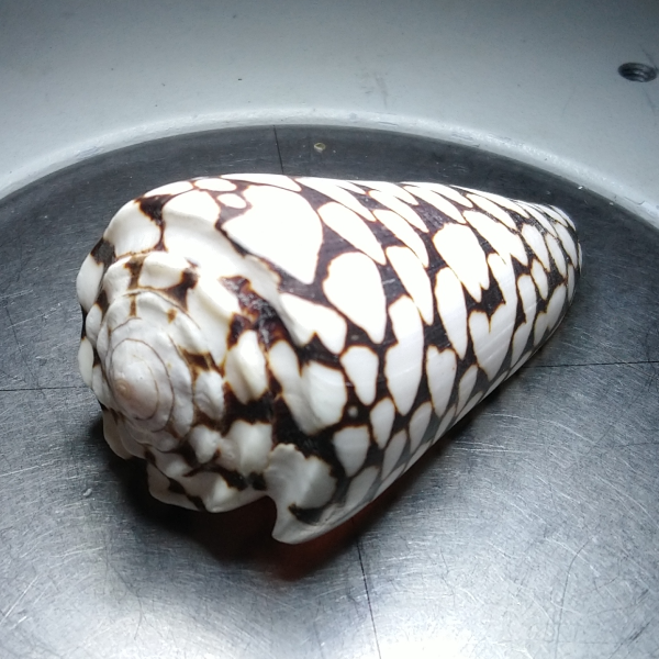
Today, I got a black cone shell which I obtained last week from a tourist shop in the north of Spain, and I think it is time I look at the other side of the shell.
A very basic description of the internal structure of the shell is that it is composed of three layers, the aragonite nacreous layer, which we have been studying before, the aragonite prismatic layer on top of it, and the periostracum or external layer.
As you can see, for sample preparation, I broke some chunks of the shell and used some Blu Tack paste to hold them to a metal plate. If you do the same, be aware that the paste can take a bit to settle down and you will have drift until it settles (maybe half a day on the worst cases), and also that it is hard to make things sit horizontal, so maybe you will need to adjust the position several times.
So... any differences between the dark and bright areas? Not that I can see (see the figure below).
Morphologically, the bright and dark areas seem the same. However, what is interesting to me is that there seem to be two different layers, one closely packed, and the other showing very clearly individual elements. My bibliographic search so far couldn't tell me what are these structures, so if you happen to know, please share your knowledge. Before zooming in to see more details, I tried digging, because the contamination and the shell should go away at different rates, but everything went away at the same speed, so it is unlikely that the structures are contamination, and it is more likely that they are part of the shell.
The figure below shows some small scan images to better see the structures, but with my lack of knowledge on these animals, I cannot identify what is what. So if you happen to know, get in touch, would love to know more.
Let's recap. We added a new structure to our shell morphology bibliotheque, the periostracum of the black cone shells. The structures we saw in this case, are similar to the ones seen in other shells? How is the internal structure of the periostracum? We don't know yet, but we do know that it is the same structure in both the dark and bright areas. How is this useful? We don't know yet either, but could help us identify other structures in the future.
I hope you find this useful, entertaining, and try it yourselves. Please let me know if you use some of this, and as usual, if you have suggestions or requests, don't hesitate to contact me.
28.10.2025
Discover the capabilities of WaveMode AFM in characterizing bottlebrush polymers with unprecedented detail and speed, ...

27.10.2025
Read this blog and discover advanced alloy engineering and cutting-edge AFM techniques for high-resolution, ...

14.10.2025
Discover how WaveMode technology resolves the tobacco mosaic virus structure under physiological conditions with ...

08.12.2024
Learn how to make a Python code to interface your AFM with a gamepad.
01.10.2024
Discover how different types of glass age and degrade over time, and learn how to use AFM technology to investigate ...
11.07.2024
FridayAFM: learn how to perform datamining on large sets of AFM data.
Interested in learning more? If you have any questions, please reach out to us, and speak to an AFM expert.