Discover the capabilities of WaveMode AFM in characterizing bottlebrush polymers with unprecedented detail and speed, ...

01.03.2024

Héctor here, your AFM expert at Nanosurf calling out for people to share their Friday afternoon experiments. Today I show a thing or two about skin, and most importantly, how to prepare skin samples for AFM.
About two years ago, having breakfast in a hotel in Switzerland, a colleague seated next to me to have his coffee (bad mistake). He had a strong background in biology, hence I started enquiring about things to image with an AFM. Among the many secrets I unveil (you see, for me they were treasures, but for most of the people with bio background probably they are common knowledge), it was the recipe to extract skin cells for AFM imaging. He said something like (and excuse my bad memory):
"get some cell tape, stick it to your skin, peel it off and you will get skin cells ready to be imaged".
... and I didn't believe it and I left the recipe archived somewhere at the bottom of my mind, thinking that it can make a nice conversation starter, but probably it will never work as he was probably oversimplifying the procedure.
Well, my apologies Patrick for not believing you. It does work, and it is quite reliable.
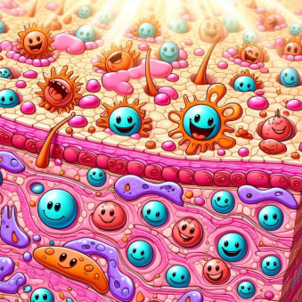
First thing to understand is that the skin is formed of many layers with different names, and when I refer to skin cells, I'm talking about the outermost layer, which is called stratum corneum, and is formed of something like 20 to 30 layers of "dead" cells called corneocytes.
Corneocytes don't have nucleus and cannot divide, but they can absorb water, hence they retain information about the status of your body when they were formed, and the things your skin has been exposed to (from UV light to drugs passing by biomarkers for cancer, see Ref 1).
Ok ok ok, all good, but can you show us how the tape stripping works? Ok, fine, here it is.
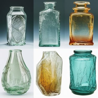
Easy enough right? What next? Well, without too much experience on this topic, It occurs to me two easy experiments. One is to compare some different cell tapes and see if there is a big difference among them (sorry, I used what I had at hand and I don't know the brands, but at least this will give you some general idea of the performance of different tapes).
It is clear that my favorite is the transparent tape. The yield is medium when compared with other two tapes, but this is good because it is possible to see both individual and clustered corneocytes, there is not too much glue residue, and for optical visualization is easy to focus and adjust the exposure and brightness. Plus, and for me with top optical microscope it makes no difference, but for those of you with an inverted system will do, the transparent tape allows to see things on an inverted system. So, this experiment was easy, transparent tape wins.
What was the other experiment? It occurred to me that maybe different parts of the body show different corneocyte morphologies. So I extracted samples from different areas and compared them. This is the map of the areas.
and these are the results.
As you can see, for both the palm (above), and the sole (below), the surface is covered with protuberances. There are likely due to wrinkles when the corneocytes shrink in size, and probably also have a function, increase the contact surface. I have no idea if the have a name.
The forearm meanwhile, presents a very different texture.
Both forearm (above), and belly (below), show a surface without protuberances (maybe the skin on the palm and sole is exposed to more friction and the skin needs to have the protuberances to guarantee mechanical stability?). It is interesting to note that while similar, the wrinkle patterns in the forearm and belly are somehow different. Apparently, the wrinkles on curved surfaces depend quite a lot on stress and stiffness, and there are phases where the pattern forms labyrinths, protuberances, or even holes (See Ref 5).
Since one of my new colleagues did a study on tattooed skin (Ref 4), and showed that the scarred tissue around the tattoo has different corneocyte morphology. I needed a tattoo. Luckily I had a volunteer (see below), but unfortunately, I couldn't locate scarred tissue, and didn't saw collagen fibers.
Last but not least, the shin where I have a mole. Seems somehow different... but it occurs to me that I should have imaged also the other shin without the mole, or a different area as a comparison. So, let's leave it that either is because of the mole that the wrinkling is here different than belly and forearm, or because this part of the skin behaves like this (again Ref 5 explains the different pattern).
Let's recap. Tape stripping is easy and produces good samples. Hopefully this tutorial has explained sufficiently how to perform the procedure. By looking at different tapes, it is clear the advantages of transparent tape (compatible with inverted microscope, medium yield allowing single cells, and good optical contrast). In terms of the skin itself, we have shown that different parts of the body are different, and the differences seem to be related with the different functionalities or environment to which that part of the skin is interacting. We couldn't see scarred tissue or collagen.
I hope you find this useful, entertaining, and try it yourselves. Please let me know if you use some of this, and as usual, if you have suggestions or requests, don't hesitate to contact me.
Further reading:
[1] Anne J. Keurentjes, Ivone Jakasa, Sanja Kezic, Research Techniques Made Simple: Stratum Corneum Tape Stripping, Journal of Investigative Dermatology,
Volume 141, Issue 5, 2021, Pages 1129-1133.e1, ISSN 0022-202X, https://doi.org/10.1016/j.jid.2021.01.004.
[2] Application note: Performing Bio-AFM on live cells
[3] Cell-cell adhesion force studied with Flex-FPM
[4] C.A. Grant, P.C. Twigg, D.J. Tobin, Static and dynamic nanomechanical properties of human skin tissue using atomic force microscopy: Effect of scarring in the upper dermis, Acta Biomaterialia, Volume 8, Issue 11, 2012, Pages 4123-4129, ISSN 1742-7061, https://doi.org/10.1016/j.actbio.2012.06.042.
[5] Stoop, N., Lagrange, R., Terwagne, D. et al. Curvature-induced symmetry breaking determines elastic surface patterns. Nature Mater 14, 337–342 (2015). https://doi.org/10.1038/nmat4202
[6] Ana S. Évora, Michael J. Adams, Simon A. Johnson, Zhibing Zhang; Corneocytes: Relationship between Structural and Biomechanical Properties. Skin Pharmacol Physiol 19 April 2021; 34 (3): 146–161. https://doi.org/10.1159/000513054
28.10.2025
Discover the capabilities of WaveMode AFM in characterizing bottlebrush polymers with unprecedented detail and speed, ...

27.10.2025
Read this blog and discover advanced alloy engineering and cutting-edge AFM techniques for high-resolution, ...

14.10.2025
Discover how WaveMode technology resolves the tobacco mosaic virus structure under physiological conditions with ...

08.12.2024
Learn how to make a Python code to interface your AFM with a gamepad.
01.10.2024
Discover how different types of glass age and degrade over time, and learn how to use AFM technology to investigate ...
11.07.2024
FridayAFM: learn how to perform datamining on large sets of AFM data.
Interested in learning more? If you have any questions, please reach out to us, and speak to an AFM expert.