FlexAFM and nanomotion sensing unveil cellular responses, offering new insights into bacterial behavior and potential ...

03.11.2025
#Done with a DriveAFM: Performance without compromise
Tobacco and tomato leaves started developing an irregular, mottled, mosaic-like pattern, leading to yield loss and stunted growth. Fruits ripened unevenly, appeared blotchy and became malformed with an altered taste. At the end of the Nineteenth century, a new plant disease emerged in Europe and America. In the 1930s, the pathogen was finally identified, and because of the patterns it caused on leaves, it was named tobacco mosaic virus (TMV). Shortly later, the American biochemist Wendell Stanley managed to crystallize it, marking the birth of molecular virology. The impact of the studies on TMV revealed the importance of investigating viral structure to understand function, and TMV went on to become a research model, valued for its exceptional stability and the high yield of its particles.
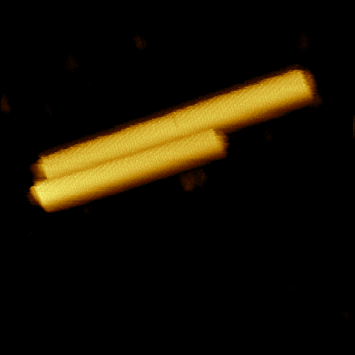
Despite all the efforts made by the community, until today observing the virus at high resolution in physiological conditions has remained an open challenge. Its cylindrical structure, 300 nm long with an outer diameter of 18 nm and a central channel of 4 nm, is wrapped within a layer of proteins, which exhibit a periodical texture with characteristic length of 2.3 nm. Now, the unique WaveMode of the Nanosurf DriveAFM can fully resolve TMV under physiological conditions without damage. WaveMode is an off-resonance atomic force microscopy (AFM) technique that unlike other implementations of off-resonance modes, has the advantage that it neither needs nor relies on subtraction of any parasitic background to maintain feedback. The WaveMode signal can also be translated into a force (in Newton), which allows fast, high-resolution imaging in liquid with a precisely defined tip-sample interaction, capturing fine structural details without compromising sample integrity.
![Tobacco mosaic virus. N:\[O]\20047 - Cantilevers for WaveMode\Measurements\WM0.1Au-SS\TMV](https://www.nanosurf.com/hs-fs/hubfs/Gallery/Tobacco%20mosaic%20virus.%20N:%5C%5BO%5D%5C20047%20-%20Cantilevers%20for%20WaveMode%5CMeasurements%5CWM0.1Au-SS%5CTMV.png?width=600&height=618&name=Tobacco%20mosaic%20virus.%20N:%5C%5BO%5D%5C20047%20-%20Cantilevers%20for%20WaveMode%5CMeasurements%5CWM0.1Au-SS%5CTMV.png)
Tobacco mosaic virus (TMV) imaged with DriveAFM in WaveMode.
Sample courtesy of Prof. Christina Wege and Ou Sha, University of Stuttgart (Germany).
Other common options for measuring the structure of small biological entities are electron microscopy or X-ray crystallography, but both are not ideal for preserving the environmental condition of a virus. Electron microscopy typically requires processing the sample with chemical fixatives, followed by dehydration and heavy-metal staining, potentially altering the virus. While being essential for stabilizing viral ultrastructure under high-vacuum and electron-beam conditions, these steps can induce conformational changes, collapse lipid envelopes, or redistribute surface proteins, thereby affecting the native morphology of TMV. X-ray crystallography of TMV necessitates crystallizing the viral particles into a highly ordered lattice, a process that inherently deviates from their native physiological state. To achieve this, TMV is typically purified and concentrated under non-physiological buffer conditions to promote crystal formation. These conditions can induce conformational constraints or restrict molecular flexibility.
In the late Nineties, researchers turned to AFM to measure the structure of TMV, but they had to perform it in dry conditions, far from native environment, or at low resolution. Later, other groups attempted to improve the measurements, but their resolution was still limited. Even if the instruments could theoretically perform better, one of the difficulties posed by TMV is its sensitivity to high loads applied by the AFM tip. A force above roughly 100 pN can cause denaturation of the virus structure, altering the sample and vanishing the efforts.
The DriveAFM built by Nanosurf offers all the solutions this problem poses. Thanks to the CleanDrive technology, the DriveAFM can excite the cantilever off resonance using an additional laser and unlocking WaveMode, the fastest off resonance AFM mode existing. Operating in WaveMode allows to accurately control the tip-sample interaction. “Resolving the molecular structure on a TMV with AFM requires superior force control and a fast feedback in liquid environment. For this, WaveMode is the solution.”, explains Paul Markus, application scientist at Nanosurf.
Being gentle on the sample is only one of the advantages of WaveMode, which is the fastest off resonance mode also in liquid. WaveMode can image up to 15 times faster than piezo-based off-resonance modes on conventional systems. Fast WaveMode imaging not only saves time, but also minimizes artifacts due to changes in conditions such evaporation of buffer or thermal fluctuations, and enables the observation of transient biological events and dynamic conformational changes.
To read more:
DriveAFM Performance without compromise
23.06.2026
FlexAFM and nanomotion sensing unveil cellular responses, offering new insights into bacterial behavior and potential ...

27.05.2026
Explore Alejandro Silhanek’s innovative spintronics research, showcasing how spin waves can be investigated leveraging ...
.jpg?width=330&height=330&length=330&upsize=true&upscale=true&name=Mayfield%20Girls-202581%20(1).jpg)
19.05.2026
A group of young girls from Mayfield school wanted to start a F24 electric car racing team, and Nanosurf decided to ...

08.12.2024
Learn how to make a Python code to interface your AFM with a gamepad.
01.10.2024
Discover how different types of glass age and degrade over time, and learn how to use AFM technology to investigate ...
02.07.2024
FridayAFM: learn how to automatize data analysis in MountainsSPIP
Interested in learning more? If you have any questions, please reach out to us, and speak to an AFM expert.