Discover the capabilities of WaveMode AFM in characterizing bottlebrush polymers with unprecedented detail and speed, ...

05.01.2024

Héctor here, your AFM expert at Nanosurf calling out for people to share their Friday afternoon experiments. Today I image my own blood.
For quite some time I've been meaning to image my own blood, but pain prevented me from voluntary extraction. This is why, the other day, when I had a little mishap trying to open an IC, I was ready to grab a glass slide and put some droplets on it.
Since it was unplanned, and my first attempt at such thing, I just put some droplets on the glass slide and let them dry, but after the experiment, I learned that usually the preparation for this type of samples involves smearing the droplets with another glass slide to create areas with different amounts of cells.
By the way, I lied, there is no graphical content here.
I had to admit that my first impression was a bit disappointed, I was expecting textbook red blood cells looking like... filled donuts? buttons? disc-shaped with thick edges... and what I got was... well, see by yourselves.
As it turns out, there are several things that can induce stress on RBCs, and these, alongside ageing of the RBCs, can modify the shape of the RBCs (See Refs 1 and 2). Apparently, the competition between the lipid bilayer and the cytoskeleton is what generates all these strange shapes.Nevertheless, why I didn't get any of the disc-shaped with thick edges?
So, first open question. Did I let it dry for too long? (About one hour), was the drying process too rough? Or because I didn't do the smearing with a glass slide I only get to see part of the RBCs population? I highly suspect is due to the sample preparation technique, because in Ref 3, they image 5300-years old blood from an iceman and looks better than mine. But the sample preparation is quite complex.
For instance, in Ref 4 they do: "The erythrocytes sample preparation was performed immediately after the blood sample collection in EDTA tubes; this was centrifuged (1000 rpm for two minutes) to discard the supernatant (plasma and buffy coat). The remaining pellet (erythrocytes) was fixed in glutaraldehyde (2.5 % in H2O, Sigma-Aldrich, St. Louis, MO, USA) and washed three times with 0.2 M phosphate-buffered saline (PBS). A drop was placed in silicon oxide (300 nm, Siltronix, Oceanside, CA, USA) and then air-dried ". This points to me that the exposure to the plasma nd the buffy coat could be the reason why in my sample there is few (if any) toroidal-shaped cells. Well, this is something to try next time around.
After looking at the shape, I tried looking at the surface of the cells (see below), because there has been publications where they study the cytoskeleton or the formation of vesicles (see Ref 5). As you can see below, I did manage to see something on top of the cells (after flattening the image quite a lot), but unfortunately, very similar granular distribution can be seen also on the substrate, meaning that this is likely residue left after the drying process (this is where the "washed three times with 0.2 M PBS" might be handy).
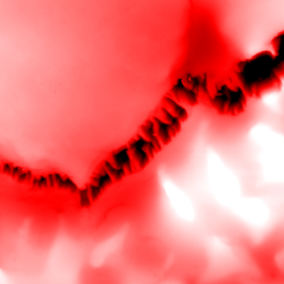
Next I noticed that around many cells there where finger-like structures. On one hand, it makes sense that they might be fibrin from the coagulation, trying to bind the cells together. On the other hand, the literature I have seen shows long filaments, not these small structures at the crevices... I have seen similar dendritic structures when salt crystals form, and I didn't prepared this sample... so there is a chance that these are just salt residues, and without further evidence to support one thing or the other, I will just report them here, hopping that in the future new data will help clarify their origin.
At this point, convinced that the RBCs were deteriorated, and that there was a layer of residue everywhere, I thought nanomechanical measurements didn't make sense, so I just finished taking a large area image to show you how small was this "usable" area of few or single-cell layer.
Let's recap. Without sample preparation (a part from letting it dry for about half an hour - one hour), we managed to image red blood cells. We saw individual cells with different shapes, likely indicating stress. Although no preparation was good enough to see cells, it was not good to see details on the surface. In addition to cells, we also saw what could be fibrin fibers, however, this last part needs to be confirmed, as salt dendrites take sometimes similar appearance.
I hope you find this useful, entertaining, and try it yourselves. Please let me know if you use some of this, and as usual, if you have suggestions or requests, don't hesitate to contact me.
Further reading:
Application note: Performing Bio-AFM on live cells.
Cell-cell adhesion force studied with Flex-FPM
[1] Lim H. W., Gerald, Wortis, Michael, Mukhopadhyay, Ranjan, Stomatocyte–discocyte–echinocyte sequence of the human red blood cell: Evidence for the bilayer– couple hypothesis from membrane mechanics. Proceedings of the National Academy of Sciences. 16766-16769, (2002). https://www.pnas.org/doi/abs/10.1073/pnas.202617299
[2] Geekiyanage, N.M., Balanant, M.A., Sauret, E., Saha, S.C., Flower, R.L., Lim, C.T., & Gu, Y.T. (2019). A coarse-grained red blood cell membrane model to study stomatocyte-discocyte-echinocyte morphologies. PLoS ONE, 14. https://doi.org/10.1371/journal.pone.0215447
[3] Janko Marek, Stark Robert W. and Zink Albert. Preservation of 5300 year old red blood cells in the Iceman. J. R. Soc. Interface.92581–2590 (2012)
http://doi.org/10.1098/rsif.2012.0174
[4] Alejandra Loyola-Leyva, Juan Pablo Loyola-Rodríguez, Yolanda Terán-Figueroa, Santiago Camacho-Lopez, Francisco Javier González, Simón Barquera, Application of atomic force microscopy to assess erythrocytes morphology in early stages of diabetes. A pilot study, Micron, Volume 141, (2021) https://doi.org/10.1016/j.micron.2020.102982
[5] Sergunova V, Leesment S, Kozlov A, Inozemtsev V, Platitsina P, Lyapunova S, Onufrievich A, Polyakov V, Sherstyukova E. Investigation of Red Blood Cells by Atomic Force Microscopy. Sensors (Basel). (2022). https://doi.org/10.3390%2Fs22052055
28.10.2025
Discover the capabilities of WaveMode AFM in characterizing bottlebrush polymers with unprecedented detail and speed, ...

27.10.2025
Read this blog and discover advanced alloy engineering and cutting-edge AFM techniques for high-resolution, ...

14.10.2025
Discover how WaveMode technology resolves the tobacco mosaic virus structure under physiological conditions with ...

08.12.2024
Learn how to make a Python code to interface your AFM with a gamepad.
01.10.2024
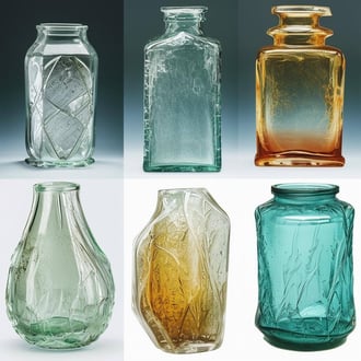
Discover how different types of glass age and degrade over time, and learn how to use AFM technology to investigate ...
11.07.2024
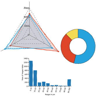
FridayAFM: learn how to perform datamining on large sets of AFM data.
Interested in learning more? If you have any questions, please reach out to us, and speak to an AFM expert.